Table of Contents >> Show >> Hide
Macular edema sounds like the kind of medical term that arrives wearing a lab coat, carrying a clipboard, and refusing to explain itself. But the idea is fairly simple: fluid builds up in the macula, the small central part of the retina that helps you read, recognize faces, drive, text, and spot the last cookie on the plate. When the macula swells, central vision can become blurry, wavy, dim, or distorted.
The tricky part is that macular edema does not always announce itself with dramatic symptoms. Some people notice straight lines bending like spaghetti. Others simply feel that reading has become harder or that one eye is “not pulling its weight.” Because the condition can be linked to diabetes, retinal vein occlusion, eye inflammation, surgery, and other retinal diseases, early diagnosis matters. The sooner a retina specialist finds the cause, the better the chances of protecting vision.
This guide explains macular edema symptoms, causes, diagnosis, treatment options, and real-life management experiences in clear, patient-friendly language.
What Is Macular Edema?
Macular edema is swelling in the macula caused by leaking fluid from damaged or abnormal blood vessels in the retina. The retina is the light-sensitive tissue at the back of the eye. Think of it as the camera sensor of your visual system. The macula is the high-definition center of that sensor. When fluid collects there, the “image quality” drops.
Macular edema is not a single disease. It is a complication or result of another problem. The most common forms include diabetic macular edema, cystoid macular edema, macular edema caused by retinal vein occlusion, and swelling related to inflammation or age-related macular degeneration.
Because the macula controls sharp central vision, macular edema usually affects tasks that require detail. Peripheral vision may stay normal, which is why some people do not realize how much central vision has changed until they cover one eye.
Common Symptoms of Macular Edema
Macular edema symptoms can range from subtle to severe. One person may notice mild blur while reading. Another may struggle to see faces clearly across a room. The symptoms often depend on how much swelling is present, how long it has been there, and whether one or both eyes are affected.
Blurred or Reduced Central Vision
The most common symptom is blurry central vision. Words on a page may look smudged. Road signs may appear less crisp. A phone screen may seem harder to read even after cleaning your glasses for the third time and blaming the poor innocent lenses.
Wavy or Distorted Lines
Macular edema can cause metamorphopsia, which means straight lines look bent, wavy, or warped. Door frames, window blinds, tile edges, or lines on an Amsler grid may appear uneven.
Colors May Look Washed Out
Some people notice reduced contrast or duller colors. Bright colors may seem less vivid in the affected eye. This can be especially noticeable when comparing one eye with the other.
Dark or Empty Spots
A person may see a gray, dark, or blank area in the center of vision. This is sometimes called a central scotoma. It may interfere with reading, recognizing faces, or seeing fine details.
Difficulty Reading or Seeing Fine Detail
Reading may take more effort. Small print may disappear in places. People sometimes describe the experience as “missing letters” or needing brighter light.
Symptoms May Be Mild at First
Macular edema can develop slowly. In diabetic macular edema, early swelling may cause no obvious symptoms. That is why regular dilated eye exams are essential for people with diabetes and anyone at risk for retinal disease.
What Causes Macular Edema?
Macular edema happens when the normal barrier that keeps fluid inside retinal blood vessels breaks down. Once the vessels become leaky, fluid seeps into the macula and causes swelling. Several conditions can trigger this process.
Diabetes and Diabetic Retinopathy
Diabetic macular edema is one of the most common and serious causes of macular swelling. Over time, high blood sugar can damage tiny retinal blood vessels. These weakened vessels may leak fluid, blood, and fatty deposits into the retina. When leakage affects the macula, vision can become blurred or distorted.
People with long-standing diabetes, poorly controlled blood sugar, high blood pressure, kidney disease, high cholesterol, or advanced diabetic retinopathy may have a higher risk. However, even people who feel perfectly fine can develop early diabetic eye disease, which is why eye exams are not optional decoration on the diabetes care checklist.
Retinal Vein Occlusion
A retinal vein occlusion occurs when a vein carrying blood away from the retina becomes blocked. Blood and fluid can back up, causing swelling and bleeding in the retina. If the macula becomes swollen, central vision may drop quickly.
Retinal vein occlusion is more common in people with high blood pressure, diabetes, glaucoma, high cholesterol, and vascular disease. It can affect a branch retinal vein or the central retinal vein.
Eye Surgery
Cystoid macular edema can sometimes occur after cataract surgery or other eye operations. This form involves cyst-like pockets of fluid in the macula. Many cases improve with treatment, especially when detected early.
Uveitis and Eye Inflammation
Inflammation inside the eye, known as uveitis, can disrupt retinal blood vessels and cause macular edema. Inflammatory causes may require anti-inflammatory treatment, steroid medications, or therapy for an underlying autoimmune or infectious condition.
Age-Related Macular Degeneration
Wet age-related macular degeneration can cause abnormal blood vessels to grow under the retina. These fragile vessels may leak fluid or blood into the macula, leading to swelling and central vision changes.
Medications and Other Eye Conditions
Some medications, inherited retinal diseases, eye tumors, trauma, and traction from the vitreous gel can also contribute to macular edema. Because causes vary, treatment should always target the underlying reason for the swelling.
How Macular Edema Is Diagnosed
Diagnosing macular edema usually involves a comprehensive eye exam and retinal imaging. A standard vision test alone is not enough because vision can remain fairly good even when early swelling is present.
Dilated Eye Exam
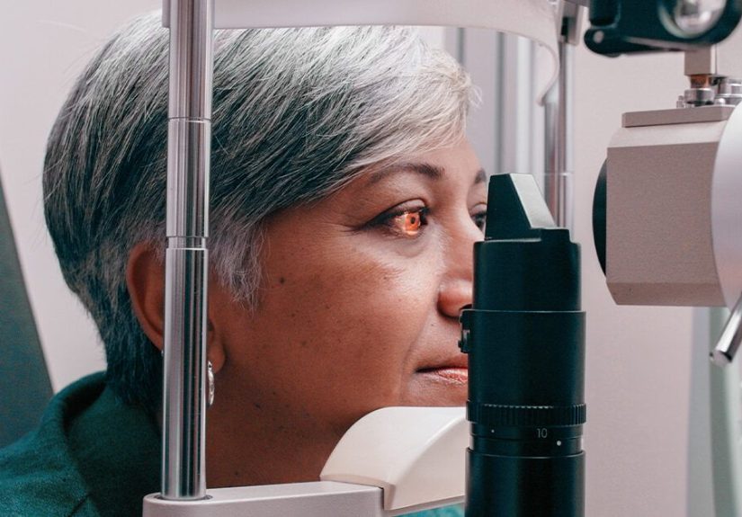
During a dilated eye exam, the eye doctor uses drops to widen the pupils and examine the retina. This allows the doctor to look for swelling, bleeding, fatty deposits, inflammation, abnormal blood vessels, or signs of diabetic retinopathy.
Optical Coherence Tomography
Optical coherence tomography, often called OCT, is one of the most important tests for macular edema. It creates detailed cross-sectional images of the retina. OCT can show retinal thickness, fluid pockets, cyst-like spaces, and changes over time. It is fast, noninvasive, and extremely useful for monitoring treatment response.
Fluorescein Angiography
Fluorescein angiography uses a special dye injected into a vein, usually in the arm. As the dye travels through retinal blood vessels, photographs reveal leakage, blocked vessels, or abnormal vessel growth. This test helps doctors identify where the leakage is coming from.
Visual Acuity and Amsler Grid Testing
Visual acuity testing measures how clearly you see letters on an eye chart. An Amsler grid may help detect distortion in central vision. Patients may also use the grid at home, but it should not replace professional exams.
Treatment Options for Macular Edema
Macular edema treatment depends on the cause, severity, symptoms, and overall eye health. The goal is to reduce swelling, stop leakage, improve or stabilize vision, and prevent further retinal damage.
Anti-VEGF Eye Injections
Anti-VEGF medications are a major treatment for diabetic macular edema, macular edema from retinal vein occlusion, and wet age-related macular degeneration. VEGF is a protein that promotes leaky and abnormal blood vessels. Anti-VEGF drugs help reduce leakage and swelling.
These medicines are delivered through an intravitreal injection, which means the medication is injected into the jelly-like center of the eye. That may sound like the plot of a horror movie, but the eye is numbed first, and many patients say the anticipation is worse than the injection itself.
Common anti-VEGF medicines include aflibercept, ranibizumab, bevacizumab, and faricimab. Some newer treatment approaches may allow longer intervals between visits for selected patients, but the schedule must be individualized.
Corticosteroid Treatments
Steroids can reduce inflammation and leakage. They may be used as eye drops, injections, or sustained-release implants depending on the cause. Steroids can be especially helpful for inflammatory macular edema or cases that do not respond well to anti-VEGF therapy.
However, steroids can increase eye pressure and raise the risk of cataract progression. For that reason, doctors monitor patients carefully.
Laser Therapy
Laser treatment may be used in certain cases, especially when leaking blood vessels are located away from the very center of the macula. Focal or grid laser can help seal leaking vessels and reduce the chance of further swelling. Today, laser is often used more selectively than in the past because injections have become a mainstay of treatment.
Anti-Inflammatory Eye Drops
For cystoid macular edema after surgery, doctors may prescribe nonsteroidal anti-inflammatory drops, steroid drops, or both. The treatment plan depends on the timing, severity, and whether other risk factors are present.
Surgery
In some cases, vitrectomy surgery may be needed. This is more likely when macular edema is related to vitreomacular traction, scar tissue, or complications such as bleeding inside the eye. Surgery is not the first choice for most people, but it can be important in selected cases.
Managing the Underlying Condition
Macular edema treatment is not just about the eye. For diabetic macular edema, controlling blood sugar, blood pressure, and cholesterol can help reduce progression. For retinal vein occlusion, vascular risk factors need attention. For inflammatory causes, the underlying inflammation must be managed. The retina may be in the eye, but it is definitely reading the body’s group chat.
Can Macular Edema Be Prevented?
Not every case can be prevented, but risk can often be reduced. Prevention focuses on protecting retinal blood vessels and catching problems early.
Get Regular Eye Exams
People with diabetes should have regular comprehensive dilated eye exams. Adults with type 2 diabetes generally need an eye exam at diagnosis because diabetes may have been present for years before detection. People with type 1 diabetes usually begin screening several years after diagnosis, according to their clinician’s recommendation. Anyone with vision changes should be checked promptly, even if the next routine exam is months away.
Control Blood Sugar, Blood Pressure, and Cholesterol
Good diabetes management can lower the risk of diabetic retinopathy and diabetic macular edema. Blood pressure control also matters because high pressure can stress fragile retinal vessels. Cholesterol management may reduce fatty deposits and vascular complications.
Do Not Ignore Vision Changes
Sudden blurry vision, new distortion, flashes, floaters, dark spots, or a curtain-like shadow should be evaluated urgently. These symptoms can signal serious retinal problems that need immediate care.
Living With Macular Edema: What Patients Often Experience
The experience of macular edema is not only medical; it is practical, emotional, and sometimes inconvenient in ways that do not show up neatly on a chart. A person may still walk around normally, cook dinner, and chat with friends, yet struggle to read a menu or see a face clearly across the table. Because central vision is affected, the condition can interfere with the little tasks that make daily life feel smooth.
One common experience is frustration with fluctuating vision. Some days may seem clearer than others. Lighting can make a big difference. A person may read comfortably in bright daylight but struggle under dim restaurant lighting. Contrast also matters. Black text on a white background may be easier than pale gray text on a glowing screen designed by someone who apparently has eagle vision.
Another experience is anxiety before treatment. Eye injections sound intimidating, especially before the first appointment. Many patients worry about pain, blinking, or moving at the wrong moment. In real clinical settings, the eye is cleaned and numbed, and the doctor uses careful technique to reduce discomfort and infection risk. Patients often report pressure, a brief pinch, watering, or scratchiness afterward rather than severe pain. Still, it is perfectly normal to feel nervous. Asking the doctor to explain each step can make the appointment feel less mysterious.
Follow-up fatigue is also real. Macular edema often requires repeated monitoring with OCT scans and multiple treatment visits. At first, appointments may be frequent. Later, the schedule may be extended if the swelling improves. Patients sometimes feel discouraged when fluid returns or when vision improves slowly. That does not always mean treatment has failed. Retinal swelling can behave like a stubborn houseguest: it may leave, come back, and require another polite but firm eviction notice.
People living with macular edema often develop helpful routines. They may check each eye separately once a week, use an Amsler grid if recommended, increase font size on devices, improve home lighting, use high-contrast settings, and keep a written list of symptoms. These simple habits make it easier to notice changes and report them accurately.
Communication with the retina specialist is important. Patients should mention new floaters, pain, redness, sudden vision loss, distortion, or missed medications. They should also share practical barriers, such as transportation problems, cost concerns, fear of injections, or difficulty attending frequent visits. Retina care works best when the treatment plan fits the person’s medical needs and real life.
Family support can make a major difference. A family member may help with transportation after dilated exams, medication reminders, blood sugar management, or simply emotional encouragement. Vision problems can feel isolating, especially when others do not notice them from the outside. A person with macular edema may look fine while silently struggling to read, drive at night, or recognize faces quickly.
The hopeful part is that many people maintain useful vision with early diagnosis and consistent care. Treatment cannot guarantee perfect results, and some vision loss may be permanent if swelling has been present for a long time. But modern imaging and therapies give doctors better tools than ever to reduce swelling, monitor progress, and personalize treatment. The key is not to “wait and see” when central vision changes. With macular edema, the smartest move is to see the right eye specialist early and keep showing up for follow-up care.
Conclusion
Macular edema is swelling of the macula, the part of the retina responsible for sharp central vision. It can cause blurry vision, wavy lines, washed-out colors, dark central spots, and trouble reading or seeing fine detail. Diabetes is a major cause, but retinal vein occlusion, eye inflammation, surgery, wet age-related macular degeneration, and other conditions can also lead to macular swelling.
Diagnosis usually involves a dilated eye exam and imaging tests such as OCT and fluorescein angiography. Treatment may include anti-VEGF injections, corticosteroids, laser therapy, anti-inflammatory drops, surgery, and management of underlying health problems. Early detection is one of the best ways to protect vision.
If you notice sudden or new changes in central vision, do not wait for your eyes to “get over it.” Eyes are wonderful, but they are not famous for sending calendar invites before trouble starts. Schedule an eye exam and get a clear answer.
Note: This article is for educational purposes only and does not replace professional medical advice, diagnosis, or treatment. Anyone with new, sudden, or worsening vision changes should contact an eye care professional promptly.